
 Librería Samer Atenea
 Kálamo Books
 Librería Elías (Asturias)
 Librería Kolima (Madrid)
 Librería Proteo (Málaga)
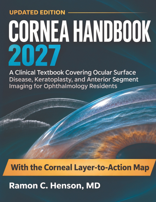
Read the Layer, Recognize the Pattern, Protect the Tissue Built for ophthalmology residents, cornea fellows, and clinicians who must convert subtle anterior-segment findings into timely action, this clinical textbook connects corneal anatomy and wound biology to diagnosis, medical care, optical rehabilitation, and surgery. It follows the full arc of practice: tear-film instability, epithelial failure, microbial and immune keratitis, dystrophies, ectasia, endothelial disease, trauma, refractive complications, keratoplasty, and lifelong graft surveillance. Layer-specific examination, slit-lamp morphology, tomography, anterior-segment OCT, specular microscopy, narrative cases, and structured decision pathways make each finding accountable to mechanism and trajectory. What This Book Puts Into Clinical Practice Localize pathology by tissue layer - distinguish epithelial, stromal, endothelial, interface, and anterior chamber processes before treatment begins Separate sight-threatening infection - integrate ulcer geography, infiltrate depth, host risk, sampling strategy, and early response Interpret shape and thickness imaging - connect topography, tomography, pachymetry, and epithelial maps to ectasia progression Restore the failing surface - manage dry eye phenotypes, neurotrophic disease, persistent defects, exposure, and medication toxicity Choose the appropriate graft - match PK, DALK, DSAEK, or DMEK to diseased tissue, recipient anatomy, and visual potential Recognize postoperative danger early - detect detachment, rejection, infection, pressure injury, suture complications, and graft failure Use the Corneal Layer-to-Action Map - move from visible signal through mechanism and pattern to an explicit action threshold and safety boundary Bring clearer structure to the next corneal finding and make every follow-up test a defensible clinical prediction.